3. The Arrival of Gene-Expression Profiling
As presented in
Table 1 and
Figure 1, several relevant DLBCL studies reported on the use of gene-expression assays with different platforms. As a cornerstone, Alizadeh et al. [
7] were the first in 2000 to demonstrate a large diversity between DLBCL cases in a microarray-based gene-expression study. This study defined two molecularly distinct DLBCL subtypes with either germinal center B-cell (GCB) or activated B-cell like (ABC) phenotypes, as shown in
Figure 2. Tumors classified as GCB showed a significantly superior overall survival (OS) compared to ABC DLBCL cases. Accordingly, Rosenwald et al. [
8] independently reported similar results with an additional third discriminating DLBCL subtype (designated as type 3), which has a similar survival rate as ABC DLBCL, and grouped together are generally referred to as non-GCB subtypes. These results were at the basis of identifying the COO to better understand lymphomagenesis. Several other studies aimed to optimize the GCB/non-GCB COO classification, explore other potential signatures for DLBCL, and validate previous findings [
6,
24,
25,
26,
27].
Figure 2. Genetic perspectives of B-cell lymphomagenesis. Under normal physiological circumstances, the germinal center is crucial for B-cell development and maturation, defining different cellular subtypes and states throughout this continuing process. DLBCL lymphomagenesis shows a GCB subtype in the earlier stages of development and an ABC subtype in later stages, representing COO classification. The COO classification is substantiated by distinct characteristic GEP and mutational profiles between GCB and ABC. This insight shows the importance of combining DNA NGS and GEP in a more multidimensional approach that improves classification and prognostication of DLBCL.
The advent of the NanoString nCounter platform optimized gene-expression analysis of FFPE samples. Scott et al. [
11] were the first to generate a COO classification using the NanoString technology in 2014. This approach utilized a targeted panel (Lymph2Cx), including 20 genes, and presented high intra-institutional concordance and overlap with microarray-based GEP. Subsequently, Dybkær et al. [
13] aimed to further subdivide the classified COO subtypes of GCB and ABC into centrocytes, centroblasts, memory B-cells and plasmablasts. This initially led to the design of a microarray-based assay called B-cell associated gene signature (BAGS) including 223 genes, demonstrating a significantly different progression-free survival (PFS) and OS between the four cellular subtypes. Subsequently, in 2018, Michaelsen et al. [
15] modified the BAGS assay to a new BAGS2CLINIC panel for the NanoString platform, including 128 genes, enabling fast and easy-to-use GEP with high overlap with the original BAGS classifier. Compared to the Lymph2Cx panel, the BAGS2CLINIC panel is more comprehensive and provides a more detailed stratification. Survival analyses using the COO assignment by BAGS2CLINIC indicated an inferior PFS and OS for the memory B-cell subtype compared to the plasmablast subtype, both originally classified as ABC subtypes. Although the centroblast and centrocyte subtypes were both classified as GCB subtypes, an inferior PFS was identified for the centroblast subtype, with no difference in OS.
In 2020, Tripodo et al. [
19] generated a spatial signature including 87 genes that discriminates between the dark and light zone of the germinal center, with similarities to COO and BAGS(2CLINIC) classifications. The subtypes identified by this panel showed prognostic significance, as a light-zone-like phenotype was associated with superior OS compared to a dark-zone-like phenotype.
With the advancing insight into COO, interest and understanding of the TME have increased. In 2005 Monti et al. [
9] analyzed transcriptional signatures in DLBCL and reported a so-called “consensus clustering” classification. This study implemented microarrays and sequential consensus cluster analysis to assess the stability of clusters in gene-expression data after different clustering methods. Three distinct DLBCL clusters were identified, two of which contained predominantly B-cell expression profiles characterized by oxidative phosphorylation and B-cell receptor/proliferation. In contrast, the third cluster was enriched for T-cell-mediated immune response and classical complement pathway and as such reflected the interaction of the microenvironment with the tumor. In contrast to COO, no correlation was found between the consensus clusters and survival [
9].
In 2008, Lenz et al. [
10,
28] identified a GCB cluster and two stromal signatures, characterized by their TME association. The first stromal signature (stromal-1) reflected the extracellular matrix and histiocyte infiltration and was associated with a favorable PFS and OS in comparison to the second stromal signature (stromal-2) which represented tumor angiogenesis. Over the following years, other independent studies investigated these stromal signatures and other biological markers for their relevance to survival, reporting similar findings [
21,
26,
29]. The identification of these stromal signatures emphasized the importance of studying the TME to improve the biological understanding of DLBCL [
10].
Carey et al. [
12] performed targeted GEP and identified a molecular classifier of MYC activity including 80 genes that stratified DLBCL patients into high- (MYC score > 0.5) and low-risk (MYC score < 0.5) groups. Patients with low MYC scores showed significantly better OS. This classification was further optimized by Ennishi et al. [
17] who generated a double-hit gene-expression signature (DHITsig) including 104 genes. DHITsig positivity was determined by overexpression of genes of high-grade B-cell lymphoma double hit or triple hit with
BCL2 translocations. DHITsig-positive cases showed strong cell-autonomous survival and proliferation signals and reduced dependence on the TME. Using this DHITsig, approximately twice as many tumors were classified as high-grade B-cell lymphoma than with conventional fluorescence in situ hybridization (FISH). PFS and OS were significantly worse in DHITsig-positive patients in comparison to DHITsig negative patients. Plaça et al. [
30] have successfully reproduced the MYC classifier of Carey et al. [
12] and the consensus clustering of Monti et al. [
9] in 175 samples of the HOVON-84 trial on a panel of 117 genes using the NanoString platform. These GEP signatures can facilitate the search for optimization of treatment algorithms, for example, which patients would benefit from the addition of lenalidomide to standard R-CHOP treatment (as described by Chamuleau et al. [
31]) or to common intensive chemotherapy regimens.
In summary, along with technological advancements over time, several GEP signatures of lymphoma cells have been identified which have significantly augmented the biological knowledge of DLBCL, distinguishing several COO and TME-based molecular subtypes with prognostic relevance.
5. The Tumor Microenvironment as Defined by Gene-Expression Profiling
The development of GEP technologies has offered the possibility to study the TME more extensively. To address various biological and clinical questions, GEP approaches have primarily focused on the role of fibroblasts, macrophages, or T cells in DLBCL lymphomagenesis. Targeted panels with probe sets covering genes encompassing discriminatory aspects of fibroblasts, macrophages, T cells, or other cells, and their activation and differentiation states have been utilized. With this TME-directed GEP strategy, several TME signatures have been defined in DLBCL, as presented in Figure 3.
Figure 3. Diversity of TME signatures in DLBCL. Several GEP signatures of lymphoma cells have been identified that have significantly augmented the biological knowledge of DLBCL. As presented, these signatures could be subdivided into three categories: tumor microenvironment, B-cell pathways, and signature assays. A relevant gene selection of potential pathways related to B-cell lymphomagenesis (purple), cell types within the TME (green), and other specific signature assays (grey) are depicted. GEP studies have demonstrated its added value in characterizing the DLBCL microenvironment and the discovery of early principles of their intriguing mechanisms. However biological issues remain, and further research is needed to determine the true clinical benefit.
Analysis of GEP data of 414 untreated DLBCL samples identified three distinct gene-expression signatures; GCB, stromal-1, and stromal-2 [
10]. The stromal-1 signature was associated with favorable survival. A computational CIBERSORT method incorporating 17 immune and stromal cytotypes into a 1028-gene matrix was applied to the previously produced data [
10,
14,
42]. This analysis revealed that a high prevalence of myofibroblasts, dendritic cells, or CD4-positive T cells was associated with superior PFS and OS as compared to an abundance of activated natural killer cells and plasma cells. Subsequently, a 45-gene set developed for NanoString-based profiling demonstrated a favorable survival of DLBCL with a high prevalence of these similar cell types [
14].
Furthermore, a lymphoma-associated macrophage interaction signature (called LAMIS, including 145 genes) was developed that specifically targets macrophages with the M2 phenotype that are immunosuppressive and promote tumor progression [
18]. High expression of the LAMIS-signature indicated poor PFS and OS, independent of COO subtype and International Prognostic Index (IPI) score [
14,
18]. Accordingly, Marcelis et al. [
43] characterized the TME of primary central nervous system lymphoma and reported that an increased M1-like/M2-like macrophage ratio was associated with superior OS. Keane et al. [
44] quantified the TME independent of the revised IPI and COO by evaluating the ratios of immune effectors with potential implications for the selection of patients in clinical trials.
Two novel corner stone studies investigated the TME through GEP and correlatedthese data with clinical and NGS data. Initially, in 2021, Kotlov et al. [20] performed clustering analysis on a large dataset retrieved from several publicly available datasets (n = 25, 4580 DLBCL cases). Based on functional gene signatures, four different cellular subtypes of the lymphoma microenvironment were clustered: germinal center-like, mesenchymal,
inflammatory, and depleted. The first cluster was characterized by germinal center features, the second cluster showed a high abundance of stromal cells and extracellular matrix pathways, the third cluster was associated with inflammatory pathways, while the depleted cluster lacked markers of these three defined GEP signatures. These lymphoma microenvironment clusters showed an impact on PFS and OS regardless of COO or genetic subtype, underlining its independent contribution to lymphomagenesis and clinical presentations. These clusters also demonstrated large similarities with previously discussed COO, consensus clustering, stromal signatures, and genetically defined entities (LymphGen profiles and C1-C5 subtypes) [4,5,20,37].
Simultaneously, as another landmark, Steen et al. reported on their so-called “Ecotyper”-algorithm that was generated for either solid tumors or lymphomas (mainly DLBCL). The landscapes of (tumor) cell states and lymphoma ecosystems were examined by means of bulk or single-cell RNA-sequencing. In this study, the B-cell states represented the previously discussed COO including GCB and ABC subtypes, as well as the subdivision into centrocytes, centroblasts, memory B-cells, and plasmablasts established by the BAGS2CLINIC. Accordingly, these B-cell states were associated with survival, corresponding to the COO or BAGS2CLINIC classification, and molecular classification with the LymphGen and C1-C5 clusters [4,37]. Furthermore, other cell-type states were identified along with a total of 9 lymphoma ecotypes that congregated multiple cell-type states and were equally associated with survival outcomes [21]. Thus, Kotlov et al. [20] and Steen et al. [21] individually identified distinct microenvironment subtypes associated with molecular profiles and survival outcomes.
As summarized in Figure 3, complementing GEP studies focusing on tumor cell subtyping and the lymphoma TME contributed to the insight that several cellular subtypes within DLBCL phenotypes were related to survival. Conclusively, an association is shown between the presence of high numbers of M2-type macrophages, natural killer cells, plasma cells, or increased angiogenesis with inferior survival. In contrast, abundant infiltration of myofibroblasts, dendritic cells, CD4-positive T cells, CD14-positive monocytes, extracellular matrix deposition and histiocytes demonstrated superior survival. However, results should be interpreted cautiously because validation studies are lacking.
The prognostic impact of the TME signatures does not answer the question of whether these microenvironmental features indicate an underlying interaction of infiltrating cells with tumor cells that promote tumor growth or, in contrast, represent an ultimate consequence of cellular damage and is thus initiated by the tumor itself. GEP studies have demonstrated their value in mapping the DLBCL microenvironment. Nevertheless, unresolved biological issues on the interaction and activation status of diverse cell types in the TME remain and further research is needed to determine the true clinical effect of the
interaction between lymphoma cells and the TME.
6. Clinical Impact and Future Perspectives of Gene-Expression Profiling Studies
As depicted in Figure 4, GEP analysis has the potential to elucidate the phenotype of the tumor, the composition of the TME, and the presence of immune surveillance mechanisms. Consequently, GEP studies have refined current DLBCL categorization towards a more biologically driven classification with different COO subtypes. Besides a more general consensus clustering, the GEP COO classification into ABC and GCB subtypes has changed the view of DLBCL’s biological behavior and is steadily regaining its place in diagnostic procedures above the surrogate Hans algorithm for COO based on immunohistochemical staining [27]. In addition, GEP COO classification is used to allocate patients with particular DLBCL subtypes to novel targeted clinical trials although a true
clinical benefit has not yet been established [16,45–47].
 Figure 4.
 Schematic overview of a multilayered research strategy. Combining targeted NGS, targeted GEP and imaging mass spectrometry allows for inclusive analysis of genotype, phenotype, TME and immune surveillance of the DLBCL. This methodology substantiates the conversion from the current approach towards a novel strategy including (1) Hans classification to COO diagnostic classification, (2) a general clinical prognostic score (International Prognostic Index) towards a biology-guided prognostication and ultimately (3) facilitating development from a one-size-fits-all R-CHOP treatment towards more precision medicine.
COO classification has been further developed into dark-zone-like and light-zone-like phenotypes or even more specific cell types, such as centrocytes, centroblasts, memory B-cells and plasmablasts, all harboring a prognostic impact. Other independently identified GEP clustering studies in DLBCL demonstrated predictive significance, such as stromal, immune-related, LAMIS, lymphoma microenvironment, “Ecotyper”, and other cellular-specific signatures (Table 1). However, despite the significant progress made in the refinement of these biological and predictive classifications, validation studies for these prognostic signatures are currently lacking and hinder direct implementation in routine clinical settings to optimize patient management and counseling. Besides that, GEP techniques are time consuming, many pathology departments are not equipped with these technologies, lack biostatistical tools needed for examining these signatures, and costs are currently not covered by most health insurances. Although optimizing the specificity of DLBCL classification is crucial to improving patient care, the challenge is to arrive at a consortium-oriented panel for discriminatory subtyping that is clinically relevant, easily accessible, has a short turnaround time, and is affordable for routine use.
GEP results can also be used to initiate a new era of therapeutic trials. Given the intermediate response to R-CHOP in DLBCL, better subtype classification of mainly high-risk DLBCL subgroups such as high-grade B-cell lymphoma could improve the effectiveness of targeted therapeutic strategies. Targeted therapies that complement or replace standard treatment have been investigated. For example, Davies et al. (2019) [16] studied the effect of adding bortezomib, a proteasome inhibitor, to R-CHOP treatment and reported no significant improvement in survival. Data retrieved from this trial, have been re-examined, identifying a DLBCL subgroup characterized by the prevalence of a distinct CD8 T-cell state that benefited from the addition of bortezomib to R-CHOP [21]. Therefore, further patient classification depending on COO status or particular signatures could improve the efficacy of bortezomib by more efficient upfront patient selection [48,49].
Kuo et al. [45] revealed that ibrutinib was less effective in ABC subtype DLBCL patients with high BCL2 expression. Wilson et al. [50] investigated this in more detail and reported that for patients of <60 years the event-free survival after treatment with ibrutinib and R-CHOP was 100% in the MCD (co-occurrence of MYD88L265P and CD79B mutations) and N1 (NOTCH1 mutations) subtype. Hartert et al. [51] described the favorable effect of adding lenalidomide to R-CHOP on event-free survival in patients with mutations in PIM1, SPEN or MYD88 or expression signatures including NF-�B, IRF4 and JAK-STAT. The addition of venetoclax to R-CHOP treatment reported worse outcomes than expected in patients overexpressing BCL2, underlining the necessity of analyzing involved pathways [52,53]. These examples show that intensifying molecular analysis is needed for the optimization of personalized treatment [54]. Consequently, this paves the way to re-evaluate previous clinical trials adopting targeted therapies, potentially providing new insights into the current conclusions.
Another important application of GEP results is in the management of resistance and efficacy of CAR T-cells and bispecific antibodies in DLBCL patients. These treatments have shown remarkable efficacy in chemorefractory DLBCL patients; however, in a significant proportion of patients, these therapies are still not effective [55–59]. Critical analysis of the TME and its influence on the effectivity of CAR T-cell and bispecific antibody therapy in DLBCL is lacking. GEP studies in DLBCL patients focusing on the TME will facilitate further evaluation of the disparate response to these novel treatments.
Kahle et al. [60] published a review on the contribution of molecular imaging to the understanding of the biology of lymphoma. Adding immunohistochemistry, proteomics, or imaging mass spectrometry to NGS and GEP data enables a multi-dimensional analysis of tumors related to the TME. For example, de Miranda et al. [61–63] performed imaging mass cytometry on tonsil and colorectal cancer tissues, thereby enhancing the understanding of the heterogeneous and intricate tumor-specific immune landscape of TME. Such molecular imaging analysis can complement current molecular evaluations in DLBCL to a more three-dimensional analysis, facilitating the identification of new mechanistic concepts. In addition, analysis of paired samples before and after an intervention will deepen the analysis of the TME, subclones and (acquired) therapeutic resistance [64]. The application of machine learning tools such as artificial intelligence will further enhance the utility of multi-omics data to better define distinct molecular and prognostic DLBCL subtypes [65].
In summary, to improve the understanding of the intricate molecular biology of DLBCL and the interaction with its TME, future studies should adopt a multi-dimensional strategy including immunohistochemistry, NGS, GEP and proteomics. Figure 4 shows such a multilayered approach and emphasizes that in addition to appropriate equipment, it also requires a diverse and well-trained team of molecular biologists, pathologists, hematologists, bioinformatics, and biostatisticians. Relevant findings of this multilayered analysis will ultimately be translated into manageable diagnostics that can be implemented by multiple medical centers.
7. Anatomical Localization and Age Matter
Together with other NGS studies, we demonstrated unique mutational profiles for DLBCL with a preferred localization, f.e., in primary central nervous system B-cell lymphoma, primary testicular lymphomas, intravascular large B-cell lymphoma, primary bone DLBCL, and primary cutaneous DLBCL leg-type [66–73]. These preferred anatomical localized DLBCLs were significantly associated with specific COO subtypes variably determined by GEP and Hans classification (Figure 5). Primary central nervous system B-cell lymphoma, primary breast DLBCL, intravascular large B-cell lymphoma, and primary testicular large B-cell lymphoma are mainly classified as ABC type lymphomas. In contrast, craniofacial, primary mediastinal (thymic) large B-cell lymphoma, primary ovarian DLBCL and primary bone DLBCL are mainly classified as GCB [67,74,75]. In addition, the Lymph3Cx GEP panel was developed as an update of the Lymph2Cx, which has distinguished primary mediastinal (thymic) large B-cell lymphoma from other DLBCL subtypes [75]. By applying a targeted NanoString panel we recently demonstrated that primary bone DLBCL mainly constitutes a centrocyte-like GCB-profile, while non-osseous DLBCL with a GCB subtype principally has a centroblast-like phenotype [67]. This conceptualizes that anatomical DLBCL localization is relevant for specific COO subtypes and even more for unique cellular phenotypes. In short, for COO subtypes, localization matters.
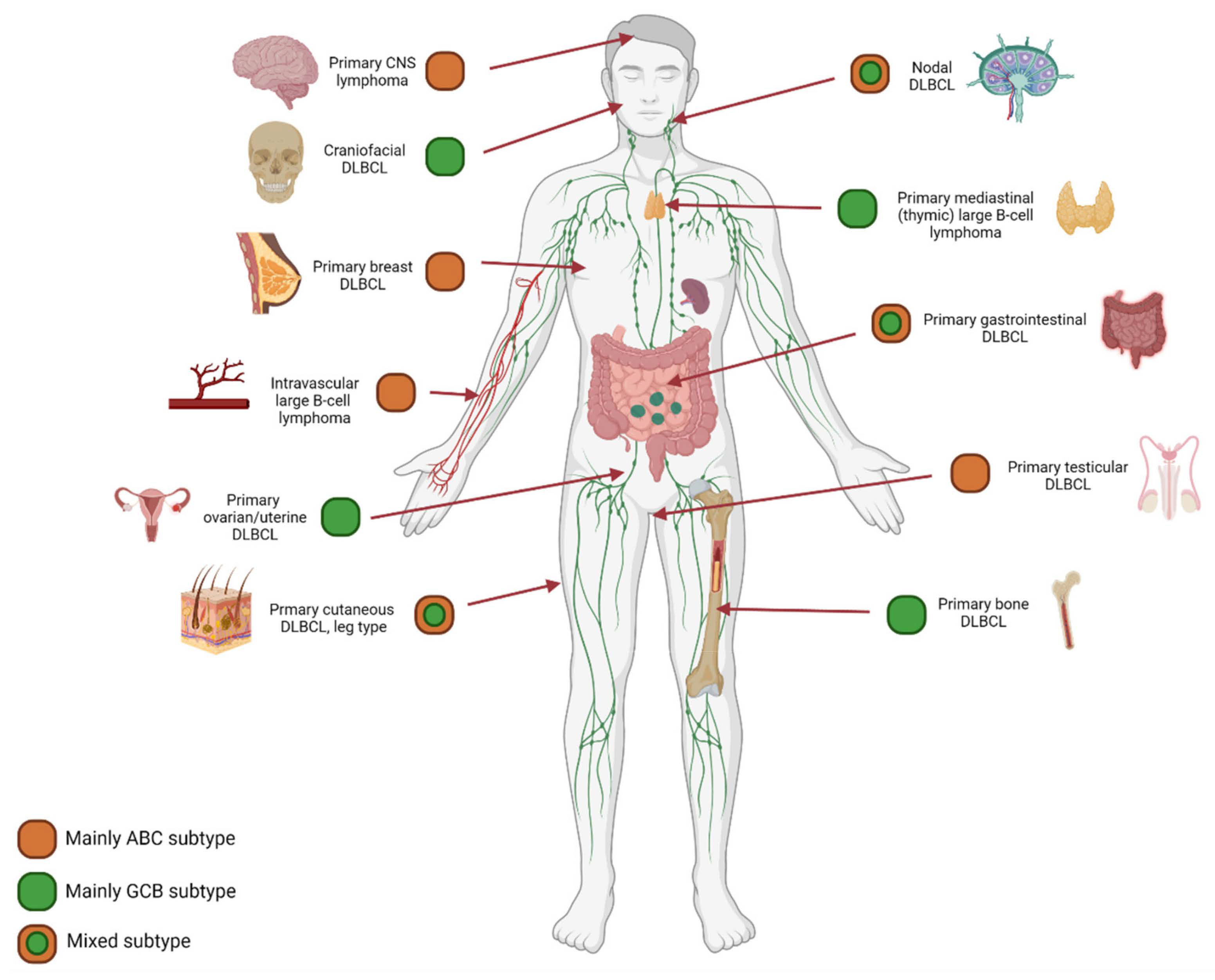
Figure 5. COO subtype: Anatomical localization matters. The results of diverse studies using GEP or Hans classification for COO determination demonstrated an evident association between anatomical preferred localization and COO subtype. For example, primary central nervous system lymphoma, primary testicular lymphoma, and intravascular large B-cell lymphoma harbor predominantly an ABC subtype. In contrast, we recently demonstrated a GCB subtype for primary bone DLBCL, that could be specified even further to unique cellular phenotypes [67]. This concept calls for additional investigation of well-annotated homogeneous cohorts of preferred localization DLBCL, including in-depth molecular studies.
Another correlation was seen between age and COO subtype, as in the elderly an ABC subtype was predominant, indicating that COO follows the physiology of senescence and alteration of the T-cell repertoire [76,77]. Altogether, these concepts have further broadened the molecular view of DLBCL, as these techniques allow COO to even be considered down to the individual cell level. As we have mentioned earlier, these results confirm the additional value of exploring well-annotated homogeneous cohorts and appeal to the need for in-depth molecular studies of DLBCL with preferred localization [78].
8. A Proposal for a Consortium Gene-Expression Profiling Panel: BLYM-777
From a biological point of view, it is important to apply GEP analysis as broadly as possible. In practice, this is often not feasible, for instance if only limited amounts of archived FFPE material are available. For this reason, a targeted GEP approach is frequently used because it is clinically applicable and allows the analysis of a limited set of genes of interest. Based on reviewing more than 45 studies and considering the maximum number of 800 genes that can be analyzed using NanoString technology, we generated a targeted knowledge-based biology-driven (t)GEP consortium panel, called BLYM-777 (Figures 3 and 6 and Appendix A). This BLYM-777 panel includes 777 genes involved in the NF-�B (f.e. MYD88, CD79B and CARD11), JAK/STAT (f.e., SOCS1, JAK1 and STAT1),
MAPK (f.e., BCL2 and MEK2), NOTCH (f.e., NOTCH3 and TBL1XR1), PI3K (f.e., PTEN and PI3K) pathways that are known to be important in lymphomagenesis of DLBCL. In addition, the BLYM-777 includes genes relevant for COO identification, such as the original COO-classification NanoString tool Lymph2Cx (n = 20 genes) (f.e., IRF4, ITPKB, MME and MYBL1), the BAGS2CLINIC (n = 53) (f.e., STAT3 and IL16), and dark-/light-zone signature (n = 52) (f.e., B2M, CTLA4, KI67 and AICDA), all of which have individually been shown to facilitate DLBCL subtype classification [11,15,19]. As the interest in the TME increases in DLBCL, BLYM-777 additionally includes genes related to TME-focused signatures, such as the consensus clustering classification (n = 86) (f.e., CD37, TNFRSF1A and PDL1), LAMIS signature (n = 63) (f.e., CCND2 and CXCR4), a 45-gene TME assay (Ciavarella et al. n = 45) (f.e., COL1A1 and MMP2), lymphoma microenvironments (n = 155) and ecotypes (n = 314) [9,14,18,20,21]. This BLYM-777 design also covers other signatures relevant for DLBCL, such as the DHITsig (n = 35) (f.e., ETV6 and RGCC) since DHIT lymphomas show an inferior survival, genes relevant in MYC driven B-cell lymphomas (n = 80) (f.e., RFC3 and TRAP1), genes upregulated in wildtype-TP53 DLBCL with high mutational burden (n = 37) (f.e., HDAC1 and BBC3), and genes relevant for the identification of resistance to CAR T-cell or bispecific antibody treatment (n = 35) (f.e., CD58 and FOXP1) [12,17,55–57,79–88]. To evaluate the influence of mutations on gene expression, 95 genes important for current molecular classification based on NGS results have also been included [67]. Supplementary Table S1: BLYM-777 included genes per author, lists all genes belonging to this BLYM-777 tGEP consortium panel. To approach multiple biologically and clinically relevant questions
on the lymphomagenesis of DLBCL, such as the relevance of certain mutations, the interaction of malignant cells with different components of the TME, immune surveillance and effectivity of new treatment strategies, the proposed BLYM-777 panel can be deployed in combination with other molecular characterization techniques. In addition, BLYM-777 is ready to use with NanoString technology and benefits from low-threshold accessibility and good performance using RNA isolated from FFPE material. However, analysis of the proposed panel of 777 genes is also possible by selective bioinformatic analysis of whole transcriptome data or targeted expression data generated by other platforms. The advantages and disadvantages of different gene-expression detection technologies have been extensively described by Narrandes et al. and Jiang et al. [22,23] and are beyond the scope of this clinical translational review.
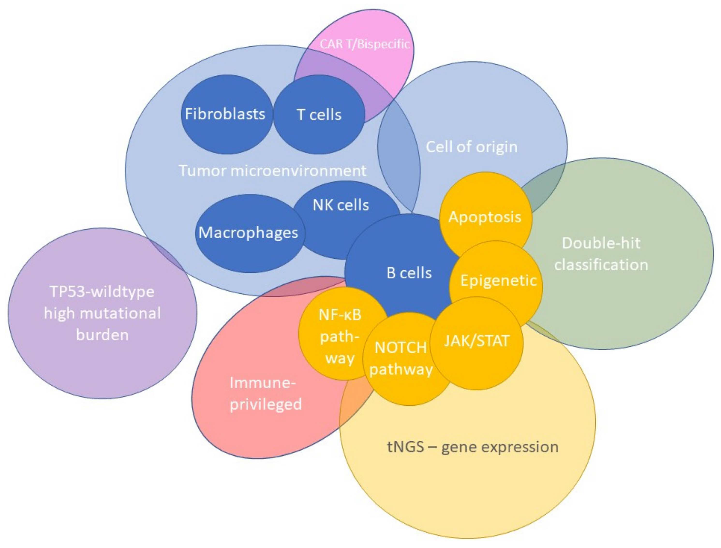
Figure 6. A proposal for a targeted BLYM-777 consortium panel. Based on 45 studies, we propose a knowledge-based, biology-driven targeted (t)GEP consortium panel, called BLYM-777. This BLYM-777 panel primarily focuses on DLBCL and covers 777 B-cell lymphoma relevant genes, including their involved pathways (f.e., NF-�B, NOTCH, PI3K). Accordingly, genes were included for COO identification, TME-focused signatures, ecotypes, DHITsig, differentially expressed genes found in wildtype-TP53 DLBCL, and genes relevant for resistance to CAR T-cell or bispecific antibody therapy. Moreover, 95 genes important for current molecular classification based on NGS results have been included.
In summary, the BLYM-777 panel covers many aspects of B-cell lymphomagenesis, COO classification, therapeutic efficacy and TME-focused signatures and can facilitate subsequent molecular investigations. The authors of this publication are currently in discussion with NanoString with the goal to create this consortium gene-expression BLYM-777 panel capturing the biology mentioned in this work. Such a panel could bring value to the hematological field by providing a standardized tool to facilitate collaboration and shared learnings throughout the community. If you are interested in joining this consortium effort, please respond to the corresponding author for more information.
9. Conclusion
This review provides a comprehensive overview of current molecular insights into the biological background of DLBCL obtained by several GEP technologies. These methods utilized in DLBCL studies identified several GEP signatures including cell-of-origin discrimination in GCB and ABC subtypes and an in-depth analysis of the TME regarding the exact cell type and state. Combining GEP with other NGS and proteomic-based methodologies will facilitate a multi-layered analysis and a next step forward in understanding biological principles and elucidating the genetic heterogeneity of DLBCL. The proposed novel knowledge-based biology-driven consortium tGEP panel, named BLYM-777, encompasses many aspects of B-cell lymphomagenesis, TME and immune surveillance and is
thereby expected to gain new molecular concepts of DLBCL lymphomagenesis. Applying BLYM-777 in such a multilayered methodology potentially enhances diagnostic classification of DLBCL subtypes, prognostication, and ultimately the development of novel targeted therapeutic strategies improving patient survival.


